Pelvic Anatomy Posterior View : Pelvis and Perineum | Radiology Key : The posterior sacrococcygeal ligament has a deep part, an extension of the posterior longitudinal ligament and a superficial part corresponding to.
byAdmin-
0
Pelvic Anatomy Posterior View : Pelvis and Perineum | Radiology Key : The posterior sacrococcygeal ligament has a deep part, an extension of the posterior longitudinal ligament and a superficial part corresponding to.. Abdominal and pelvic anatomy encompasses the anatomy of all structures of the abdominal and pelvic cavities. It can be divided into the greater pelvis and the lesser pelvis. You've got the upper region, the superior part of the pelvic going back to the ischium, if you remember the lateral view, the anteroinferior part is the pubis. Sagittal view of the pelvic organs depicting the retropubic, vesicovaginal, rectovaginal, and retrorectal spaces. The posterior sacrococcygeal ligament has a deep part, an extension of the posterior longitudinal ligament and a superficial part corresponding to.
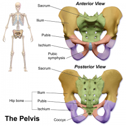
Pelvic skeleton includes two hip bones, sacrum and coccyx. Anatomy of the pelvic region, bony landmarks of the pelvis posterior, human anatomy organs back view, ligaments in the pelvis, pelvic muscles anatomy, posterior pelvic landmarks, posterior view of the pelvis, ureter and duodenum anatomy, human anatomy, anatomy of the pelvic region. But understanding this level of skeletal anatomy will make it easier to understand muscle and how can you quickly visually assess a client's propensity towards a pelvic tilt? Abdominal and pelvic anatomy encompasses the anatomy of all structures of the abdominal and pelvic cavities. This anatomy section promotes the use of the terminologia anatomica, the international standard of anatomical nomenclature.
Pelvic Floor Anatomy - Physiopedia from www.physio-pedia.com • ilium • ischium • pubis the sacrum and coccyx • form the posterior wall of the bony pelvis functions: The pelvic floor is primarily made up of thick skeletal muscles along with nearby ligaments and fascia. Agreements & disagreements workshop 36. View of the pelvic inlet and pelvic muscles from above. Posterior abdominal wall and pelvis. Dorsally, there are the posterior abdominal muscles, the back muscles, and the lumbar spine. The pelvis consists of the sacrum, the coccyx, the ischium, the ilium, and the pubis. Sagittal view of the pelvic organs depicting the retropubic, vesicovaginal, rectovaginal, and retrorectal spaces.
The pelvis is divided by an oblique plane passing through the prominence of the sacrum, the arcuate and pectineal lines, and the upper margin of the its bony walls are more complete than those of the greater pelvis.
Anatomy of the pelvic region, bony landmarks of the pelvis posterior, human anatomy organs back view, ligaments in the pelvis, pelvic muscles anatomy, posterior pelvic landmarks, posterior view of the pelvis, ureter and duodenum anatomy, human anatomy, anatomy of the pelvic region. View of the pelvic inlet and pelvic muscles from above. Anatomy of the pelvis includes anatomy of the bony pelvis and its contents. Of female pelvic organ support, with 5,6. The pelvis is separated into two regions. It can be divided into the greater pelvis and the lesser pelvis. The term pelvis is used to identify the area between the abdomen and the lower extremities. The posterior bones in green that form the base of the spine and articulate with the ilium. The lower posterior part of the abdominal and pelvic cavities the lumbar and sacral (lumbosaral) nerve plexuses exiting the… Arrangement of the flight muscles (a) cross section through the sternum (b) lateral view. Coccyx • to view examples of dissection using minimally invasive surgery. The superior surface of the bladder is. The bony pelvis & gender differences in pelvic anatomy.
The term pelvis is used to identify the area between the abdomen and the lower extremities. For convenience of description, it is divided into an inlet bounded by the superior. ƒ iliolumbar ƒ lateral sacral ƒ superior gluteal. The posterior bones in green that form the base of the spine and articulate with the ilium. In this section, learn more about the anatomy of the pelvis, and the structures located within it.
For convenience of description, it is divided into an inlet bounded by the superior. Dorsally, there are the posterior abdominal muscles, the back muscles, and the lumbar spine. Pelvic sidewall anatomy and retroperitoneal spaces. Sagittal view of the pelvic organs depicting the retropubic, vesicovaginal, rectovaginal, and retrorectal spaces. You've got the upper region, the superior part of the pelvic going back to the ischium, if you remember the lateral view, the anteroinferior part is the pubis.
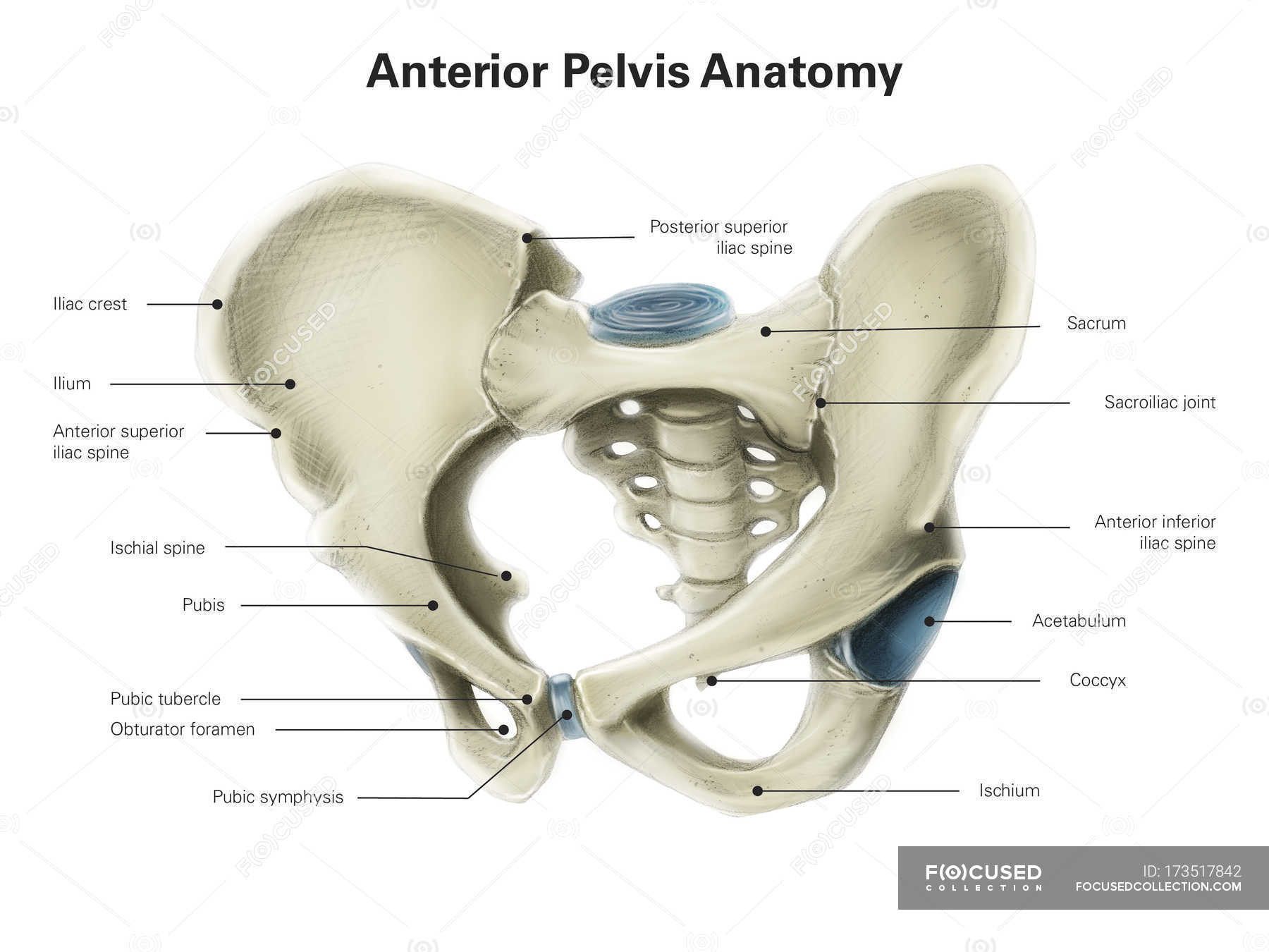
Anterior view of human pelvis — biology, human body parts ... from st.focusedcollection.com Posterior abdominal wall and pelvis. Half of this bone is part of the pubis and the other half. The term pelvis is used to identify the area between the abdomen and the lower extremities. Pelvic surgery requires a comprehensive knowledge of the pelvic anatomy to safely attain access, maximize exposure, ensure hemostasis, and avoid injury to viscera, blood vessels, and nerves. This anatomy section promotes the use of the terminologia anatomica, the international standard of anatomical nomenclature. Safe access to retroperitoneal structures. Contemporary views on female pelvic anatomy. Note the gender difference in distance between both cirsta iliaca anterior superior (distantia interspinosa), the.
The pelvis consists of the sacrum, the coccyx, the ischium, the ilium, and the pubis.
The geometry of bony pelvis front view of the male and female pelvis. It can be divided into the greater pelvis and the lesser pelvis. Contemporary views on female pelvic anatomy. Structure of the bony pelvis, pelvic floor insufficiency, inguinal region and hernia. It can help you understand our world more detailed and specific. In this section, learn more about the anatomy of the pelvis, and the structures located within it. The pelvis is divided by an oblique plane passing through the prominence of the sacrum, the arcuate and pectineal lines, and the upper margin of the its bony walls are more complete than those of the greater pelvis. Vides a discussion of the contemporary understanding. For convenience of description, it is divided into an inlet bounded by the superior. The posterior sacrococcygeal ligament has a deep part, an extension of the posterior longitudinal ligament and a superficial part corresponding to. We hope you will use this picture in the study and. Bony pelvis or pelvic skeleton is formed by hip bones, sacrum, and coccyx. From a lateral view, assess.
From a lateral view, assess pelvic anatomy. Anatomynote.com found pelvic region posterior view from plenty of anatomical pictures on the internet.